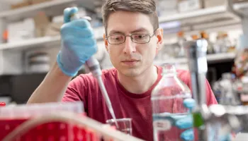
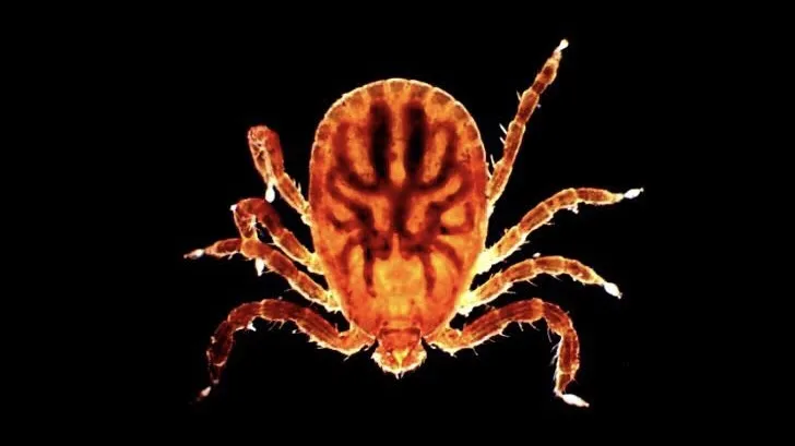
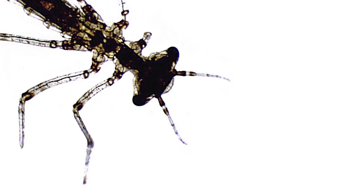
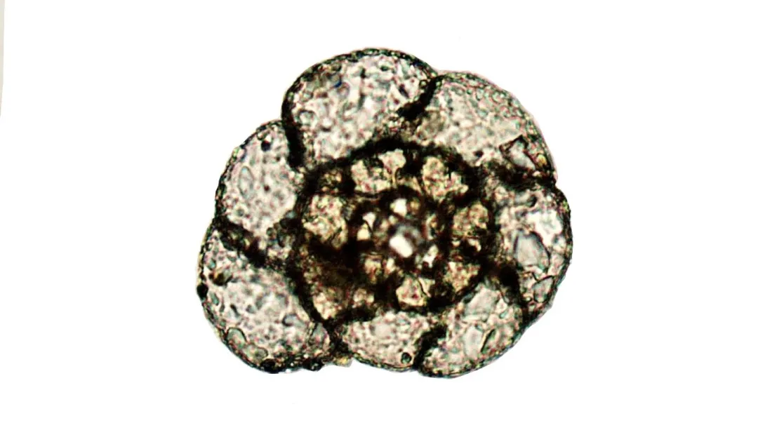
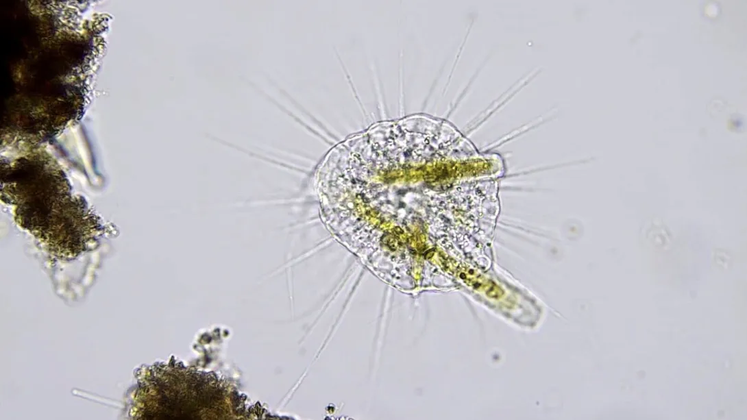
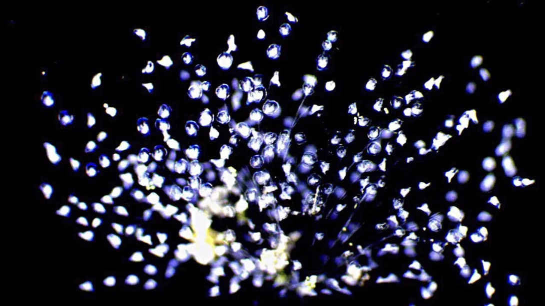
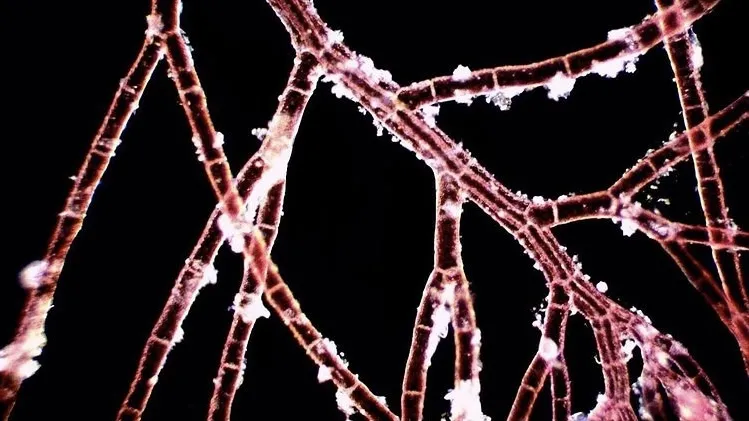
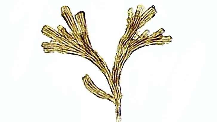
Assistant Professor Julia Van Etten researches how organisms acquire genes from other species through a process called horizontal gene transfer.
Julia Van Etten didn’t expect to make one of her biggest discoveries while on vacation. The assistant professor of biology who joined the University of Maryland this summer was in the Outer Banks, North Carolina, with her family when she passed a roadside marsh, grabbed a water sample and brought it back to her rental house.
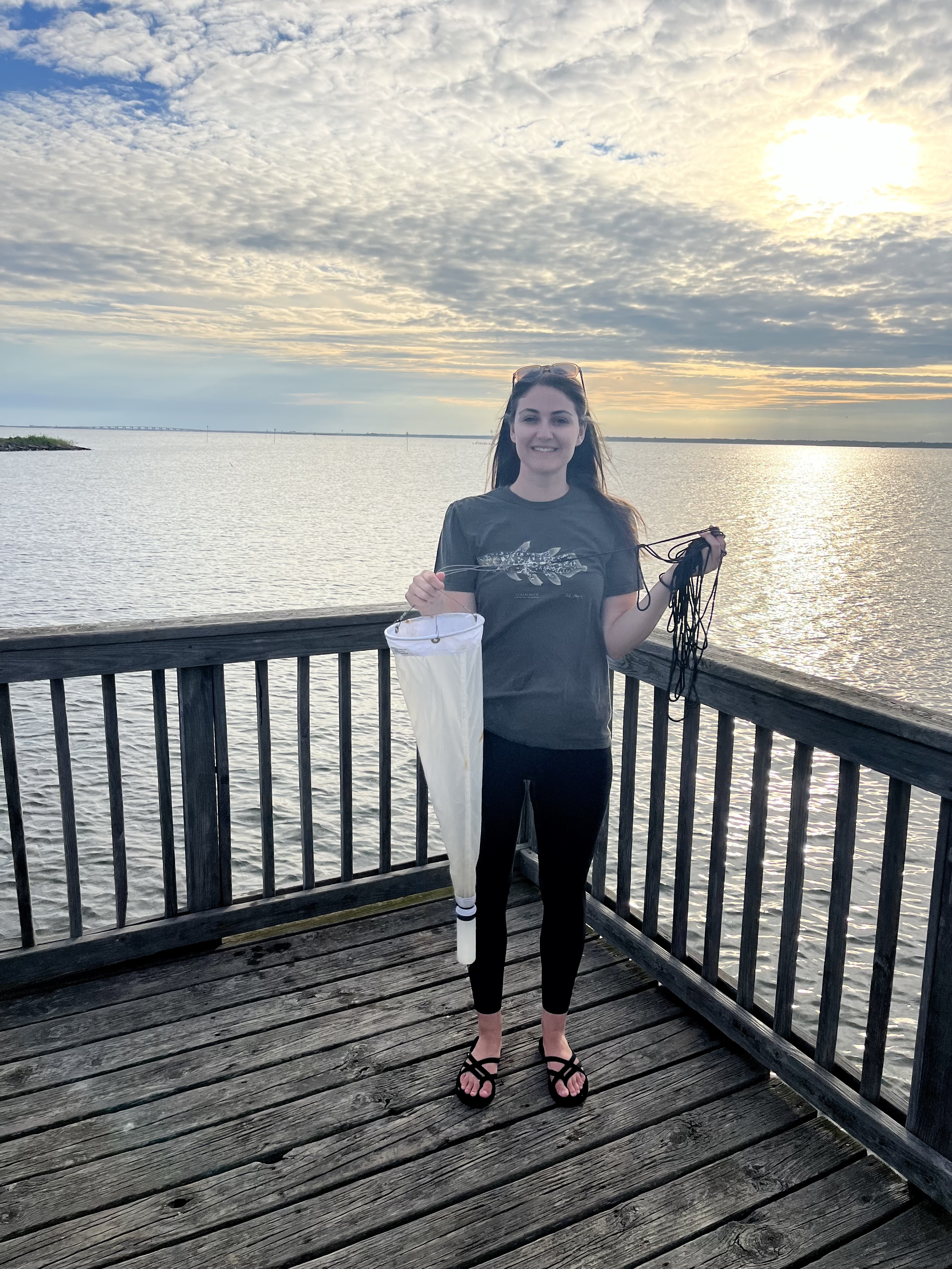
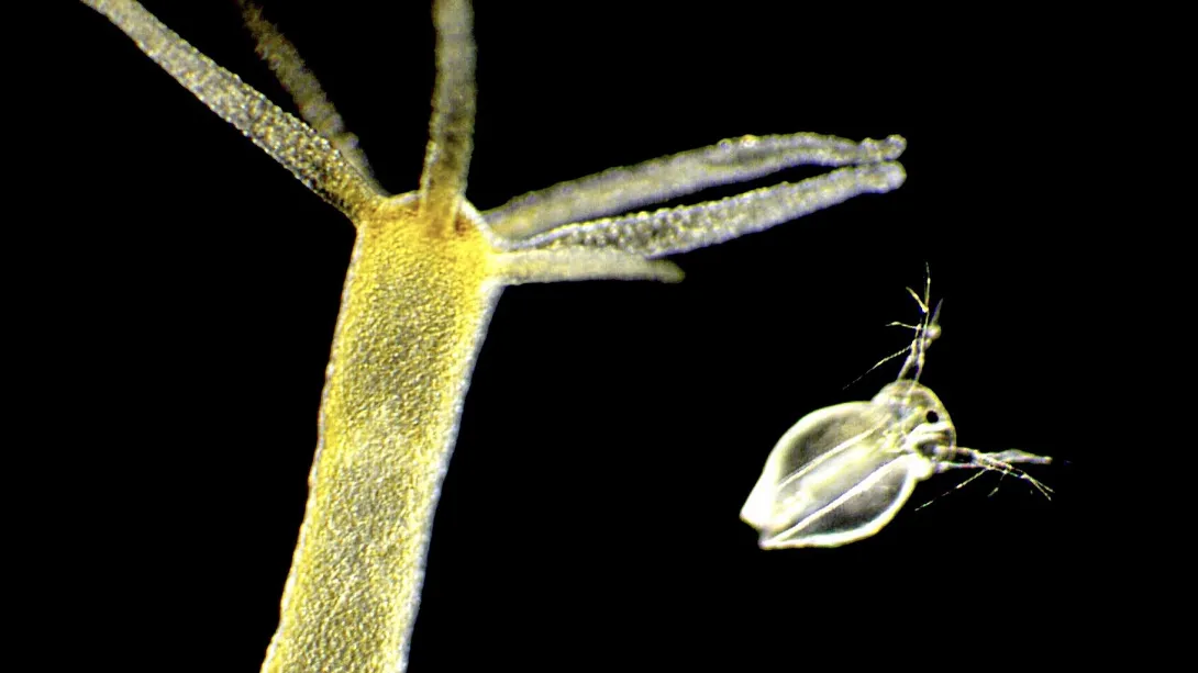
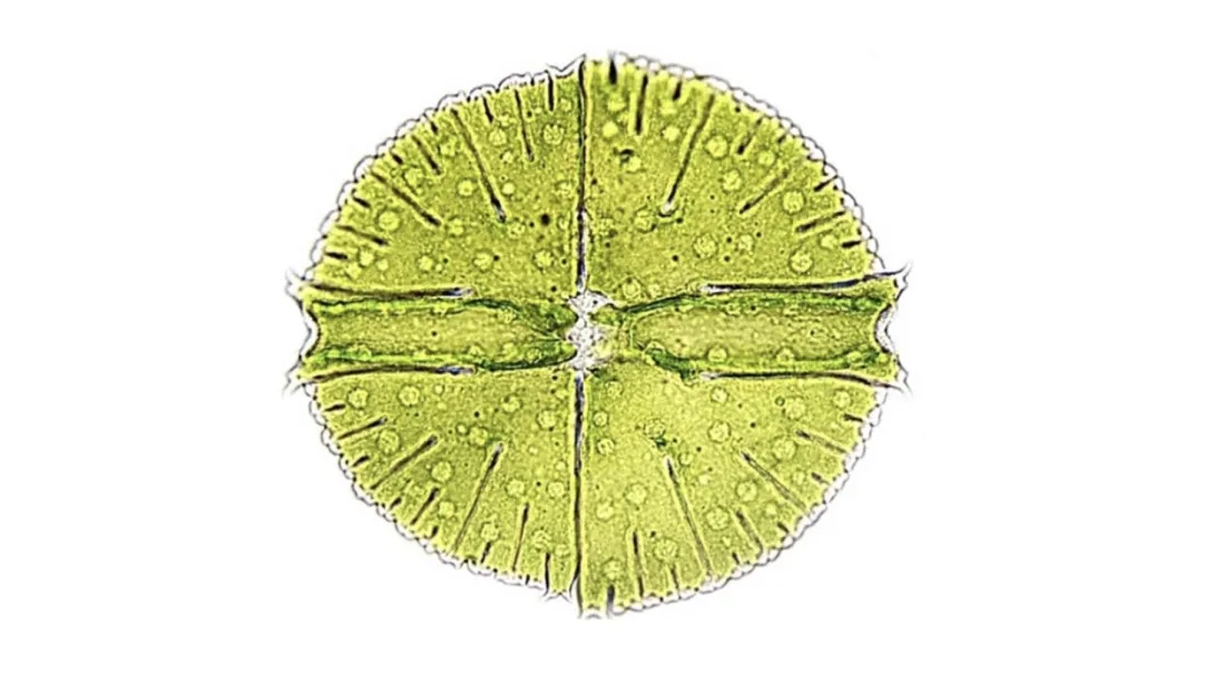
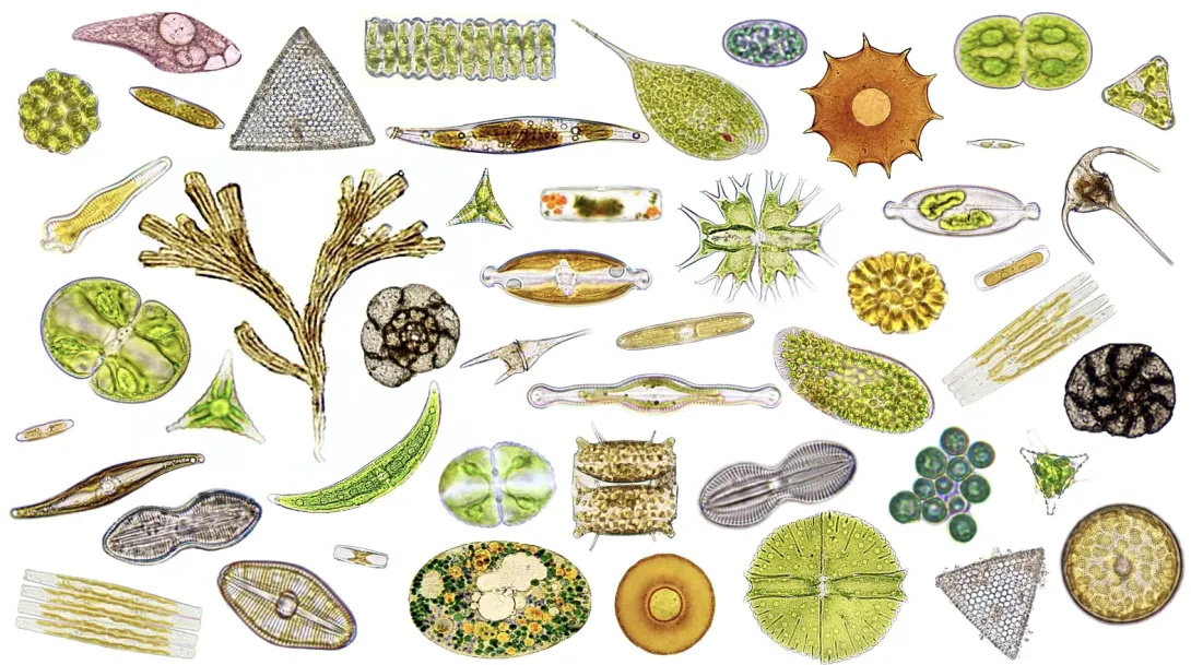
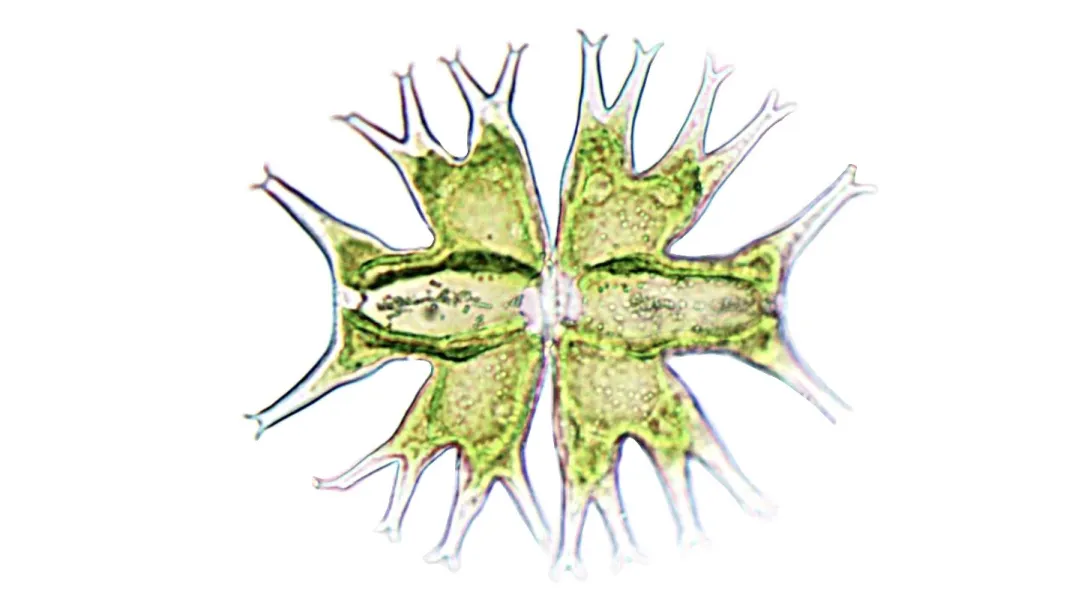
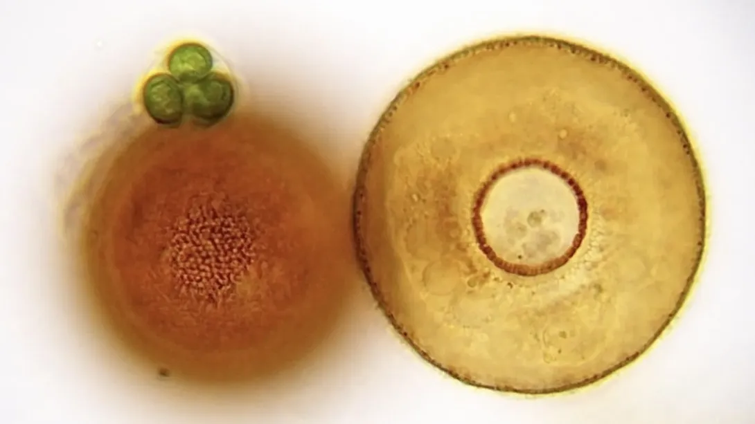
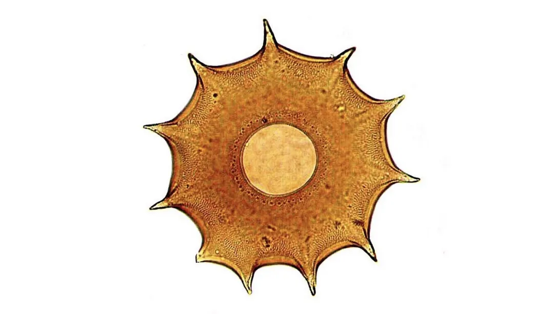
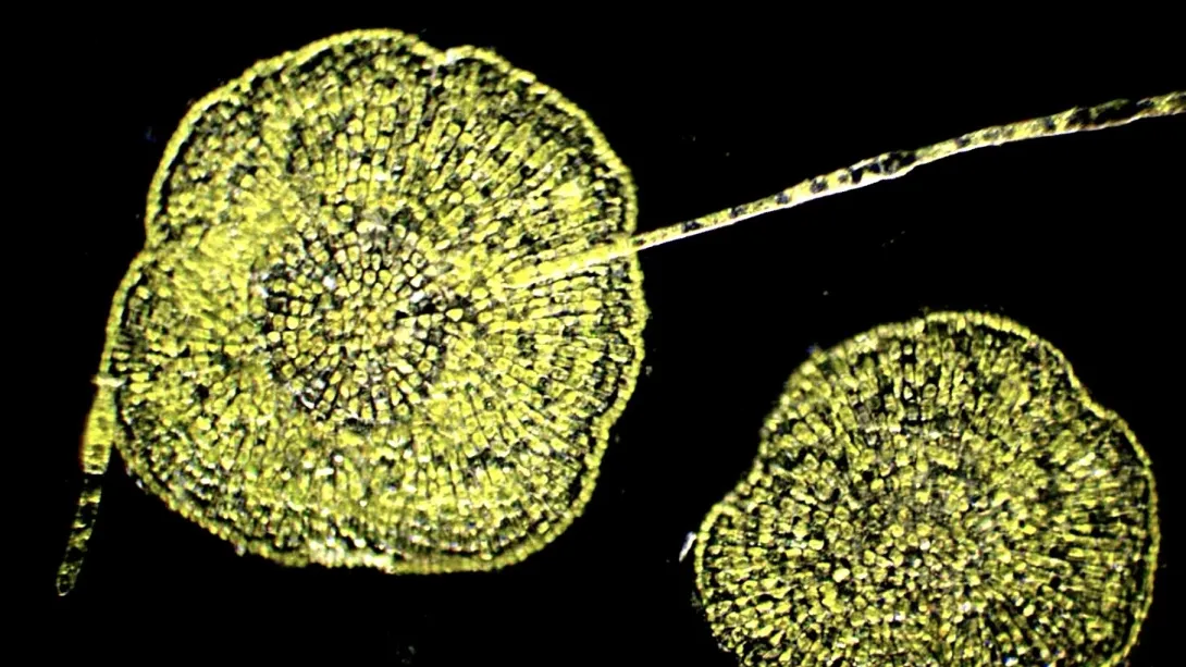
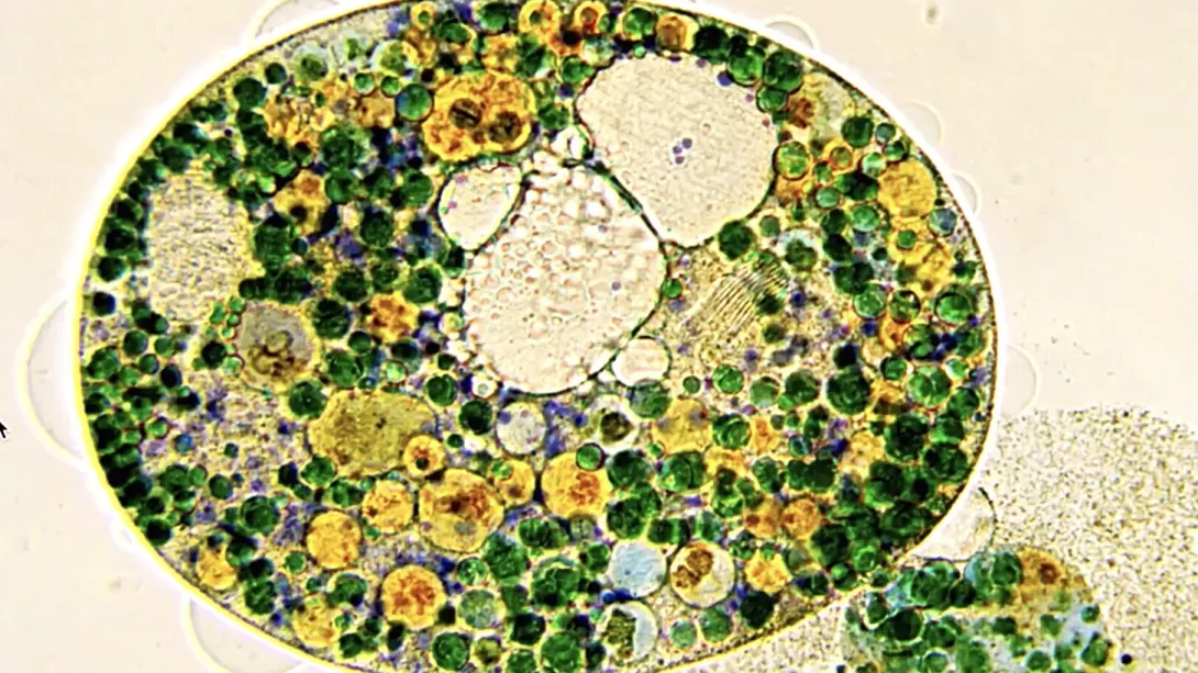
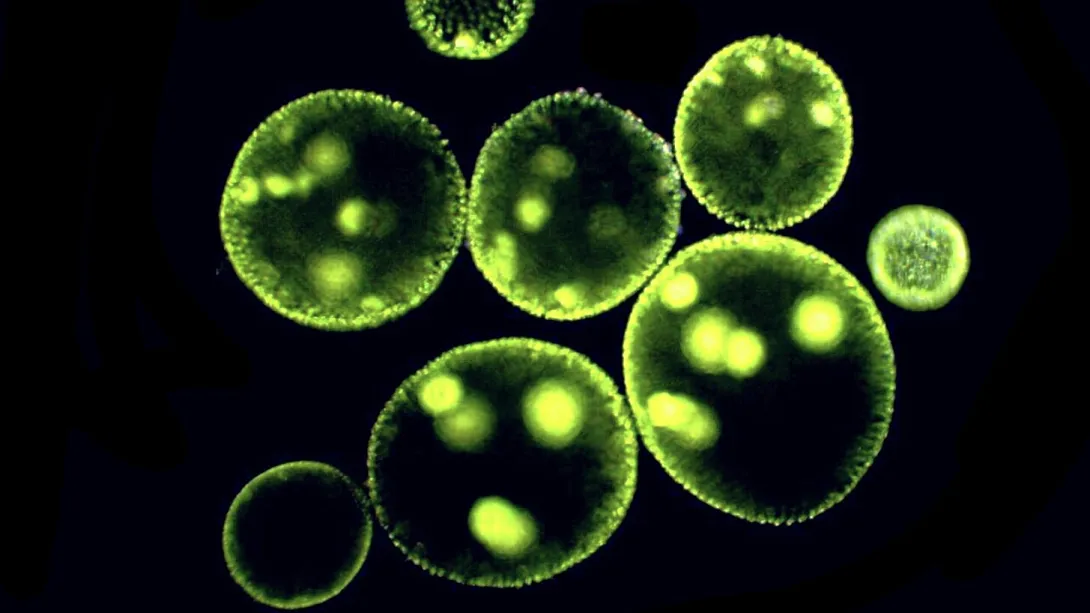
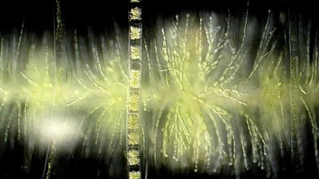
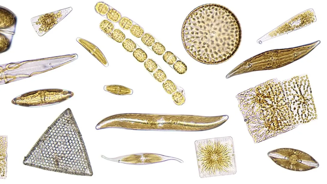
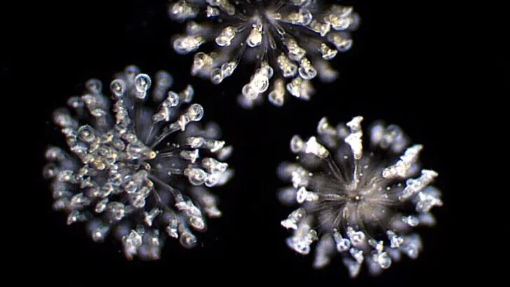
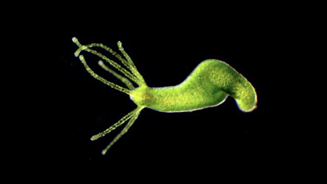
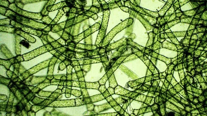
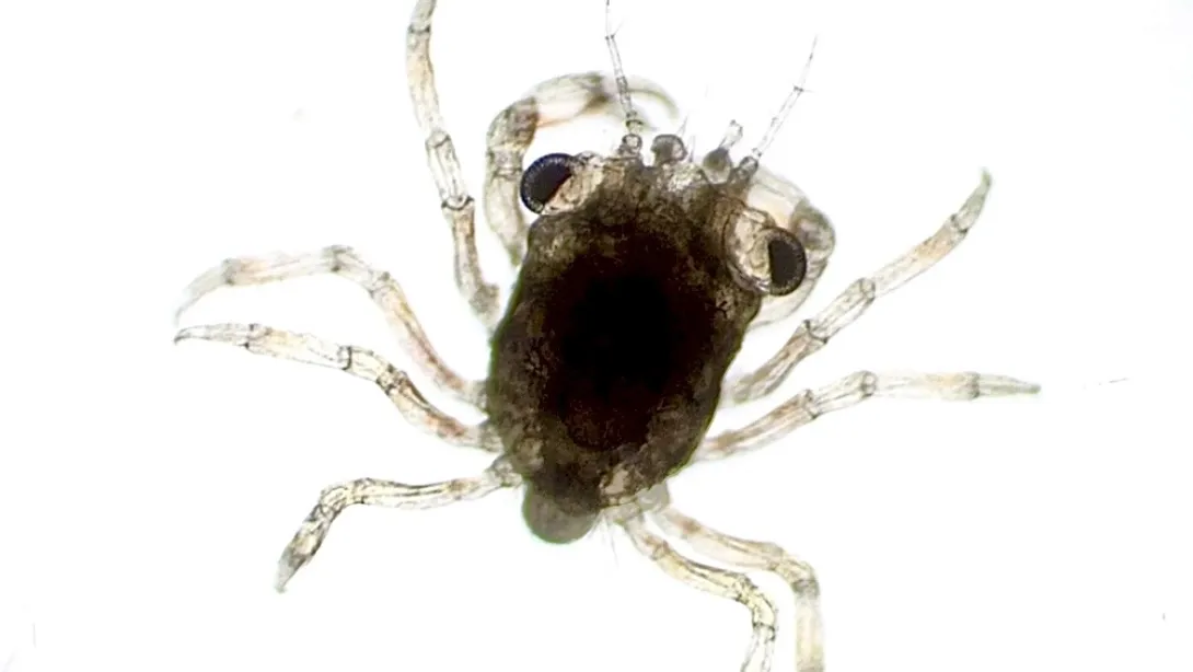
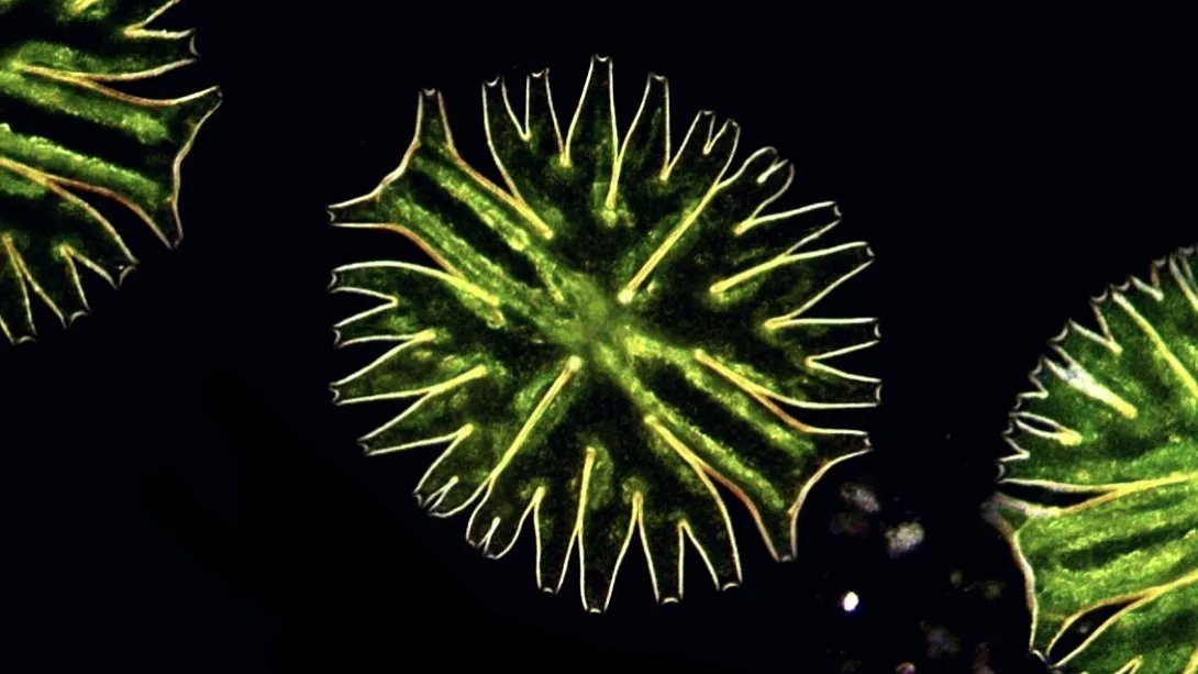
There, Van Etten pulled out the hobby-grade $300 microscope and $198 camera she packed—necessary equipment for her art and science communication project, Couch Microscopy, where she photographs and video records overlooked microorganisms people encounter in their everyday lives. Among other species, she’s visualized swimming pink protists, roving marine worms and triangular phytoplankton called diatoms. Her Instagram account, which she started before graduate school, now has more than 30,000 followers.
“I don't think that you have to go to the ends of the Earth or to exotic locations to find cool life. I get the most joy out of finding interesting-looking things and interesting-behaving organisms in very ordinary locations,” Van Etten said. And for her, sampling that Outer Banks marsh was nothing out of the ordinary.
Van Etten often dips her net into ponds, puddles and other quotidian bodies of water. But when she got back to her room and looked at this sample under the microscope, she saw two cells she’d never seen before.
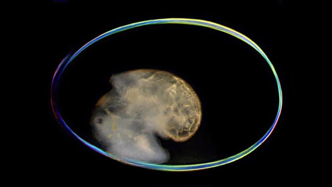
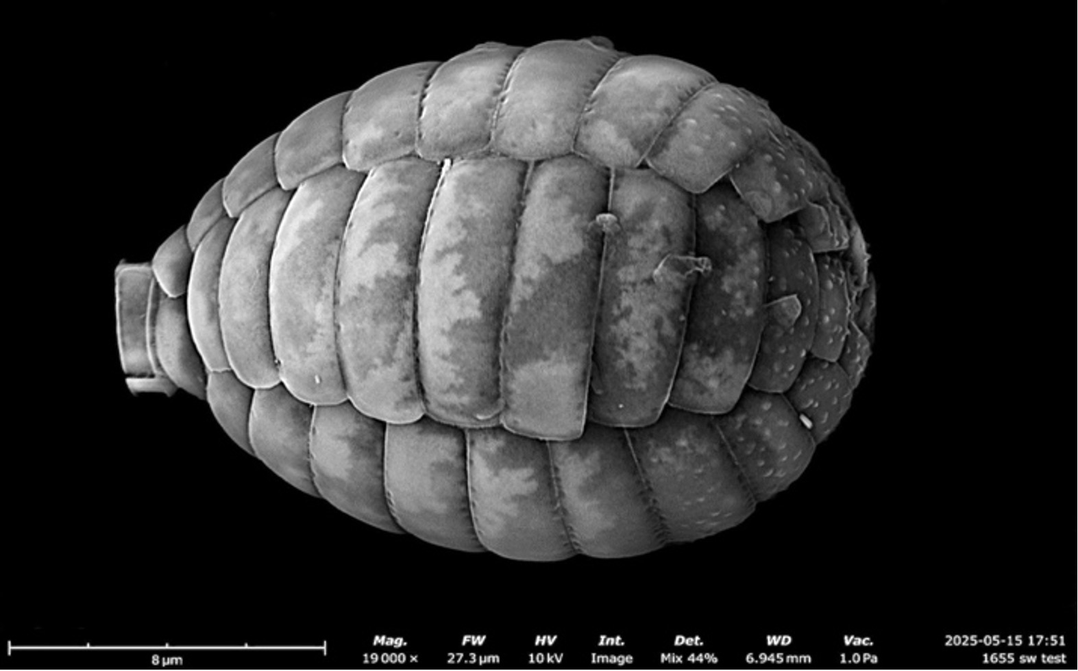
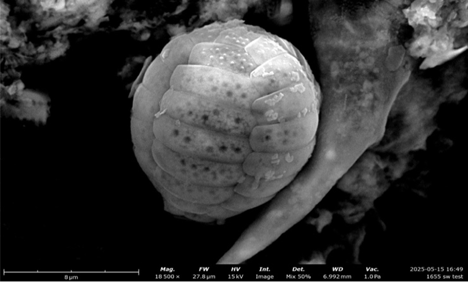
She realized they belonged to a rare group of amoebas called Paulinella. Although each cell was only about 15 microns in length, they looked distinct enough that she realized they were two different, undescribed species—and they could be the key to advancing her research program. At UMD, she studies how organisms acquire new DNA and organelles through so-called “horizontal processes,” in which they steal traits from other organisms in the community rather than inherit them from their parents.
“I believe horizontal processes are some of the most fundamental genomic processes that have shaped evolution, ecology, life on Earth and the biodiversity that we see today,” Van Etten said.
Horizontal gene transfer: how organisms steal DNA
The process by which an organism takes DNA from another species and embeds it into its genome is called horizontal gene transfer (HGT). This can happen in several ways—for example, when a cell picks up stray fragments of DNA from the environment, eats another organism or is infected by a virus.
It had long been known that bacteria and archaea transfer DNA through HGT, but it wasn’t until modern genome sequencing efforts that scientists broadly accepted that HGT plays an important role in the evolution of more complex organisms like plants, animals, fungi and protists. These organisms, called eukaryotes, have their DNA bound in a membrane, so there’s a barrier for foreign genetic material to embed itself into the genome.
During Van Etten’s Ph.D. research at Rutgers University, she studied HGT in a group of single-celled red algae called Cyanidiophyceae. The species she worked on live in the hot springs of Yellowstone National Park. Despite their name, Cyanidiophyceae grow in bright green mats because they lost the ability to produce the pigment that gives their relatives a warm red color. It’s in part because of HGTs that these red algae can thrive in Yellowstone’s scorching temperatures, acidity and heavy metal concentrations.
Van Etten’s research validated that certain species of Cyanidiophyceae acquired genes from neighboring bacteria that help them detoxify metals like arsenic. She also showed that HGT is happening in the community in real time. Today, the organisms in the ecosystem each perform different tasks that contribute to neutralizing the environment of heavy metals. In a sense, Van Etten says, DNA can be a public good, transferred between these organisms for collective survival.
Over the past few years, Van Etten scaled up this research as a National Science Foundation Postdoctoral Research Fellow in Biology at the Woods Hole Oceanographic Institution, where she studies HGT in the Narrow River in Rhode Island. This six-mile-long river has a gradient of oxygen levels, where the top is loaded with oxygen and the bottom layer has none. Van Etten studies how DNA is transferred within and between layers.
It’s not just organisms in niche, specific habitats that have benefited from HGT, either. Studies show that the process could have played a critical role in the evolution of the first plants and animals, allowing them to acquire new traits that aided in their success in new and changing environments. Much like how the even earlier acquisition of bacteria that became mitochondria and chloroplasts led to the origin of eukaryotes and algae respectively, these events paved the way for the most complex life we see today.
Van Etten hopes to probe how new organelles evolve by studying the two species of Paulinella that she discovered on vacation. These amoebas recently evolved an organelle that allows them to photosynthesize, similar to a chloroplast. HGT appears to have helped integrate the new organelle into its host’s biology, and since this resembles what happened when mitochondria and chloroplasts first evolved billions of years ago, studying Paulinella could be scientists’ best chance to gain firsthand knowledge of some of the most consequential events in evolution.
Paulinella: a model for primary endosymbiosis
A hundred million years ago, an ancestor of Paulinella ate a cyanobacterium and turned it into a new photosynthetic organelle called a chromatophore. This process, called primary endosymbiosis, is similar to how mitochondria and chloroplasts evolved in two separate events about 2 billion years ago. It’s a horizontal process, similar to HGT, but at a larger and more permanent scale. Those three instances, plus a fourth event involved in the evolution of a new organelle discovered in a marine alga in 2024, are the only times that scientists know primary endosymbiosis happened, Van Etten said.
Van Etten and her colleagues have long wanted to study Paulinella to understand how primary endosymbiosis happened and how it shaped early eukaryote evolution. The issue was that few scientists knew where to collect Paulinella or how to sustain them in the lab. That’s where Van Etten’s vacation discovery comes in.
In a paper published September 4 in the Journal of Phycology, Van Etten described the two new species she discovered, named Paulinella marae and Paulinella murrayi, after family members who were with her and encouraged her to sample from the location where she first collected the species. She is starting lab cultures of the two species at UMD. If she’s successful, she’ll move on to cutting-edge genomic and transcriptomic studies to understand how the new organelle evolved—including the role that HGT played.
Van Etten is also engaging other hobby microscopists and natural historians who have spotted Paulinella around the world, creating a new consortium that she hopes will have the collective knowledge to sample and describe countless new species. Eventually, she wants to understand how the ability to photosynthesize changed Paulinella genomes and ecological roles across their evolutionary tree.
This two-pronged approach, combining cutting-edge lab techniques with old-school naturalism, is at the heart of Van Etten’s research program. Her work has greatly benefited from the hours she’s spent staring at local microorganisms through her microscope.
“The best way to learn about nature is to be in it,” she said, noting that she learned more during her first few weeks sampling for Couch Microscopy than she had in her 22 years of life before starting the account.
Based on her large Instagram following, Van Etten suspects this sort of backyard naturalism attracts an audience for science communication, too.
“I think people really like the concept that they could walk by a puddle, and there’s all this life in it that they can’t see,” she said.
So, she’ll train her students to think like backyard natural historians. She wants to equip her lab with an arsenal of hobby-grade microscopes her trainees can use to explore. If they’re interested, she’ll even help them develop their own science communication platforms.
“It’s good for the soul to just go out in nature and see what happens,” Van Etten said. “The closer you are to your system, the better you understand it, and the more creative you’ll get with your questions and insights—and that leads to more success in science.”